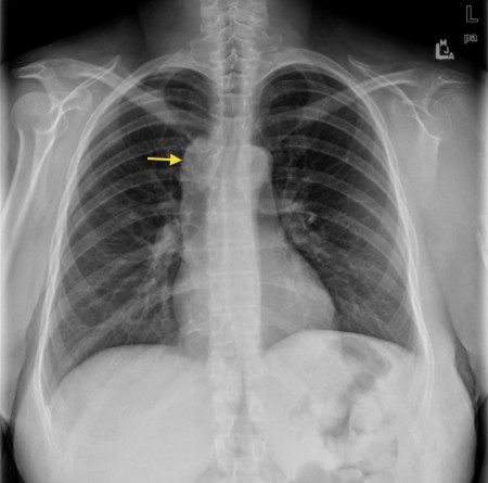
The sensitivity of ultrasound for detecting nodes of more than 1 cm is 16.7% higher than clinical examination. Lymphadenopathy in the supraclavicular region can be difficult to detect on clinical examination. 4 Historically, there has been knowledge of occult supraclavicular lymph node involvement which may be sampled surgically. 3 Cytological aspiration of palpable nodes is a well established technique. Supraclavicular lymph node involvement is considered N3 disease according to the TNM classification. 2 A rapid relatively non-invasive test would therefore be beneficial. Survival rates for advanced lung cancer are poor, being 8.4% for stage III disease and only 1.6% for stage IV. 1 Patients being considered for palliative and radical treatment need both accurate staging and cell typing as prognosis and treatment are related to both. It is estimated that, of those patients with newly diagnosed lung cancer, 26% will have involved mediastinal nodes and 49% distant metastases at presentation. Recent 2002 statistics revealed that lung cancer accounts for 33 600 deaths per year, representing 22% of all cancer related deaths in the UK. Lung cancer is a common disease with over 38 410 new cases diagnosed in the UK in 2000. As a result of FNAC, 43 patients (42.6%) avoided more invasive procedures.Ĭonclusion: Ultrasound guided FNAC is a promising, relatively non-invasive technique for the staging and diagnosis of patients with lung cancer. The overall malignant yield was 45.5% of patients scanned and 75.4% of patients sampled. Results: Sixty one of the 101 patients had enlarged supraclavicular nodes and underwent FNAC. FNAC was performed on all supraclavicular nodes over 5 mm in size using the capillary aspiration technique. Methods: 101 patients were enrolled prospectively over a 1 year period. If positive, this technique helps to both stage the patient and provide a cytological diagnosis. Ultrasound of the neck with fine needle aspiration cytology (FNAC) of enlarged but impalpable supraclavicular nodes has been used in patients with suspected lung cancer who have N2 or N3 disease on staging computed tomography (CT). Pathological diagnosis traditionally requires invasive procedures such as bronchoscopy, mediastinoscopy, or image guided biopsy.


mesentary metastasis of carcinoid tumor ( C7B.04).malignant neoplasm of lymph nodes, specified as primary ( C81- C86, C88, C96.-).Malignant neoplasms of ectopic tissue are to be coded to the site mentioned, e.g., ectopic pancreatic malignant neoplasms are coded to pancreas, unspecified ( C25.9).For multiple neoplasms of the same site that are not contiguous, such as tumors in different quadrants of the same breast, codes for each site should be assigned. 8 ('overlapping lesion'), unless the combination is specifically indexed elsewhere. A primary malignant neoplasm that overlaps two or more contiguous (next to each other) sites should be classified to the subcategory/code.Primary malignant neoplasms overlapping site boundaries.In a few cases, such as for malignant melanoma and certain neuroendocrine tumors, the morphology (histologic type) is included in the category and codes. The Table of Neoplasms should be used to identify the correct topography code. Chapter 2 classifies neoplasms primarily by site (topography), with broad groupings for behavior, malignant, in situ, benign, etc.
#SUPRACLAVICULAR LYMPH NODES CODE#
An additional code from Chapter 4 may be used, to identify functional activity associated with any neoplasm. All neoplasms are classified in this chapter, whether they are functionally active or not.


 0 kommentar(er)
